
What Does a Knee Replacement Look Like on an X-Ray?
If you've recently had knee replacement surgery, your first postoperative X-ray might surprise you.
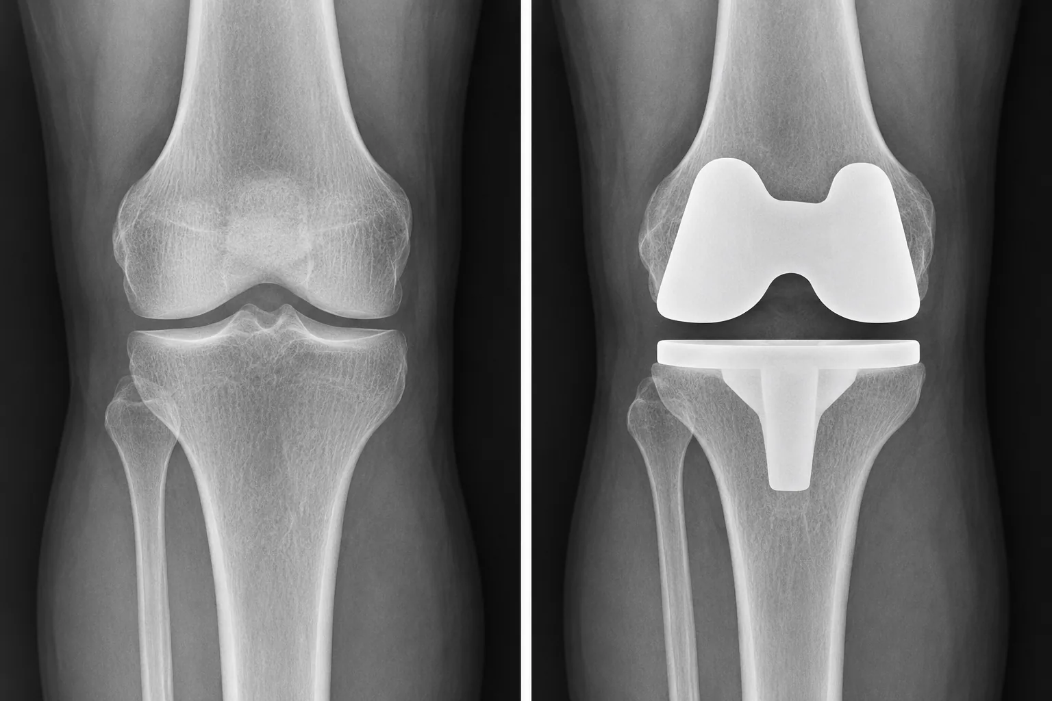
Instead of the familiar outline of a natural knee, you'll see bright metal shapes at the ends of two bones. There's a darker space between them, and your kneecap still appears at the front.
You might wonder: Where is the plastic part? Is that gap normal? And how can the surgeon tell whether everything is in the right position?
A knee replacement X-ray can answer some of these questions. It shows the metal components of the artificial joint and how they sit against the surrounding bone. The plastic bearing surface, however, is usually not directly visible.
Once you know what you're looking at, the image becomes much easier to understand.
What Does a Knee Replacement X-Ray Actually Show?
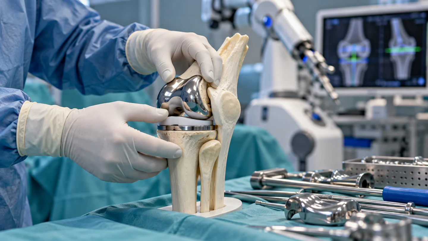
A total knee replacement usually appears as two bright metal components with a darker space between them. The curved upper component covers the prepared end of the thighbone, while a flatter component sits on top of the shinbone. A plastic bearing surface separates them, although ordinary X-rays generally do not show the plastic directly.
Here's what you can identify:
- The curved metal part: The femoral component, attached to the thighbone.
- The flatter metal part: The tibial component, attached to the shinbone.
- The darker space: The region containing the polyethylene plastic insert.
- The bone at the front: The kneecap, or patella, which may or may not have been resurfaced.
These components replace the damaged joint surfaces, not the entire thighbone or shinbone.
If you're considering surgery, understanding how robotic assistance supports knee replacement planning may also help explain how implant positioning and knee alignment are assessed during the procedure.
The implant itself doesn't necessarily look different because robotic technology was used.
Start With the Curved Metal Part at the Top
On a side-view X-ray, one of the easiest parts to recognize is the curved metal component at the lower end of the thighbone.
This is called the femoral component.
Your natural thighbone ends in two rounded surfaces that help the knee bend and straighten. When arthritis damages the cartilage covering those surfaces, movement can become painful and restricted.
During total knee replacement, the surgeon removes damaged cartilage and a measured amount of underlying bone. A specially shaped metal component is then fitted onto the prepared surface.
From the side, the implant often looks like a smooth metallic curve.
From the front, its appearance is different. You may see two rounded metal contours corresponding to the reconstructed joint surfaces.
That's why the same replacement can look quite different depending on the X-ray angle.
The metal component is designed to move against a plastic bearing surface when the knee bends. Its exact shape and size depend on the implant system and the patient's anatomy.
What Is the Flat Metal Piece on the Shinbone?
Look beneath the curved femoral component.
You'll usually see a flatter metal structure sitting across the upper end of the shinbone.
This is the tibial component, often called the tibial tray or baseplate.
It supports the plastic insert and transfers loads into the shinbone when you stand or walk.
Some tibial components have a short stem or keel extending into the bone. Others use different fixation designs.
The appearance can vary considerably between implant systems.
A patient may notice what looks like a small metal projection extending down from the tray and assume something unusual has happened. In many designs, this is simply part of the implant's normal structure.
The surgeon interprets these features according to the particular prosthesis used.
A single image without that context may be misleading.
Why Is There a Dark Gap Between the Metal Parts?
This is often the most confusing feature of a knee replacement X-ray.
The thighbone has a metal covering. The shinbone has a metal platform. Yet there appears to be an empty space between them.
That space normally contains the artificial bearing surface.
The insert is usually made from polyethylene, a durable medical-grade plastic designed for joint replacement.
Unlike metal, polyethylene absorbs relatively few X-rays. It therefore appears much less distinctly on a conventional radiograph.
The metal components are bright, while the plastic between them is generally invisible.
That contrast creates the impression of a gap.
What does the plastic insert actually do?
It provides a surface on which the metal femoral component can move as the knee bends and straightens.
In a natural knee, cartilage covers the ends of the bones and helps them move smoothly.
During replacement, damaged joint surfaces are reconstructed using artificial materials.
The polyethylene insert forms an essential part of that new bearing arrangement.
Its shape and thickness vary according to the implant design and surgical requirements.
A darker gap on an X-ray does not, by itself, mean the plastic is missing, worn out or incorrectly positioned.
Those concerns require interpretation by the treating surgeon, sometimes using previous images or additional investigations.
What Happens to the Kneecap on an X-Ray?
Your kneecap remains at the front of the knee after a routine total knee replacement.
On a side-view radiograph, it can often be seen in front of the metal femoral component.
During surgery, the surgeon may retain the natural surface underneath the kneecap or resurface it with a plastic component.
Resurfacing does not involve removing the entire kneecap. A portion of its underside is prepared to receive an artificial bearing surface.
Because that component is usually plastic, it may be difficult to identify directly on an ordinary X-ray.
Some patellar implant designs contain features that are visible on radiographs.
When the surgeon needs a closer assessment of the kneecap's position, additional views may be requested.
The decision to resurface the patella also varies according to the patient's clinical circumstances and the surgical approach.
So if two knee replacement X-rays show slightly different appearances around the kneecap, that does not automatically indicate a problem.
Normal Knee vs Replaced Knee: What Changes?
A natural knee and a replaced knee can look remarkably different on an X-ray.
Before surgery, the image mainly shows the bones, their alignment and the apparent space between their joint surfaces.
Cartilage itself is not directly visible on an ordinary X-ray.
Doctors may assess changes in joint-space width, bone spurs and other findings when evaluating arthritis.
After total knee replacement, bright metal components become the most noticeable structures.
The NHS explanation of how knee replacement is performed describes the use of metal and plastic components to reconstruct the damaged surfaces of the thighbone and shinbone.
An X-ray lets you see much of this reconstruction, although it cannot reveal every implant material or surrounding soft tissue.
Why Do Doctors Take X-Rays From Different Angles?
Imagine looking at a chair directly from the front. You can judge its width, but you cannot easily see how far the seat extends backwards.
A side view gives you different information.
The same principle applies to knee X-rays.
A single view cannot show every aspect of implant position.
Front-view X-ray
The front view, often called an AP or anteroposterior X-ray, helps the surgeon examine the relationship between the metal components and the bones.
It may help assess component positioning, certain alignment features and the bone around the implants.
Weight-bearing images can provide additional information in selected circumstances.
Side-view X-ray
The lateral or side-view X-ray shows the curved femoral component particularly clearly.
It also helps the surgeon assess the tibial component's position, its relationship with the shinbone and the appearance of the kneecap.
Full-length standing X-ray
Sometimes, an image extending from the hip to the ankle is useful.
This helps assess the alignment of the entire lower limb rather than only the knee joint.
Such views may be relevant during surgical planning or selected postoperative assessments.
Not everyone requires all these images. The appropriate views depend on the procedure, symptoms and clinical findings.
Can an X-Ray Show Whether the New Knee Is Straight?
X-rays help surgeons assess component position and alignment.
But the goal is not simply to make every implant appear perfectly straight in an isolated picture.
A knee replacement must function during walking, bending, standing and other movements. Its overall alignment is interpreted alongside the patient's anatomy and the surgical plan.
Doctors may assess the orientation of the components relative to the thighbone and shinbone. In some situations, they also evaluate the mechanical alignment of the entire leg.
These measurements are more involved than drawing a line through the implant on a single image.
Two patients can have different anatomical characteristics and still receive appropriately planned replacements.
HealMyBones also discusses this subject in its article on why knee replacement alignment is planned individually.
That distinction matters because an X-ray can help evaluate implant position, but it cannot tell you whether the ligaments feel balanced or whether the knee is stable throughout its movement.
Can You Tell From an X-Ray Whether Robotic Surgery Was Used?
Usually, no.
Robotic assistance describes technology that supports aspects of the operation, not necessarily a different type of implant.
Depending on the system, it may assist the surgeon with anatomical measurements, surgical planning, positioning guidance or selected operative steps.
The resulting metal and plastic components may resemble those used in knee replacements performed without robotic assistance.
A postoperative X-ray therefore cannot reliably establish which surgical technology was used.
That information should come from the operative record and the treating team.
Robotic technology also does not eliminate the need for follow-up imaging, rehabilitation or clinical assessment.
For someone comparing surgical options, the more useful discussion concerns why a particular approach and implant have been recommended for their knee.
Can an X-Ray Reveal Problems With a Knee Replacement?
An X-ray can identify certain changes that deserve further assessment.
For example, doctors may examine the position of the implants, changes in the surrounding bone and areas that could suggest loosening or wear-related problems.
One finding that sometimes appears in reports is a radiolucent line.
This means an area looks darker near the interface between the implant and bone or cement.
Such a line does not automatically mean that the replacement has become loose.
The surgeon may consider its position, size, whether it is changing over time and whether the patient has symptoms.
Comparing current images with earlier X-rays can be particularly helpful.
The American Academy of Orthopaedic Surgeons' patient-facing joint replacement report describes implant-related complications, including mechanical loosening, that may sometimes require further treatment.
Still, an X-ray has limits.
It does not directly show muscle strength, pain levels or every ligament and soft-tissue problem. It also cannot reliably exclude infection.
If a patient has persistent or worsening pain, the orthopaedic team may need a physical examination, blood tests or other investigations.
An X-ray finding should not be interpreted in isolation.
Why Are Follow-Up X-Rays Needed?
An early postoperative X-ray can document the initial appearance and position of the replacement components.
That image may become useful later if the surgeon needs to compare implant position or investigate new symptoms.
Follow-up imaging can help identify changes over time, but the schedule is not identical for every patient.
The surgeon considers the implant, symptoms, medical history and clinical findings before deciding when radiographs are required.
More imaging is not automatically better.
X-rays use ionising radiation, so each examination should have a clear clinical purpose.
If your surgeon recommends another image, it is reasonable to understand what information it is expected to provide.
When Should You Discuss Your Knee X-Ray With an Orthopaedic Surgeon?
You don't need to understand every measurement on an X-ray report.
But you should be able to understand the main findings and how they relate to your recovery.
After knee replacement, increasing pain, repeated giving-way, worsening stiffness or reduced ability to walk deserves assessment.
Fever, wound drainage, increasing redness or significant new swelling may indicate a complication requiring prompt medical attention.
Dr. Manoj Kumar Khemani provides orthopaedic and joint replacement consultations in Kolkata through HealMyBones.
During an appropriate consultation, the imaging can be reviewed alongside the patient's symptoms, knee movement, stability and rehabilitation progress.
For patients searching for a knee replacement surgeon in Kolkata, clear communication about surgical planning and postoperative assessment is an important part of informed care.
The X-ray is useful, but the patient's actual knee function remains central to the clinical assessment.
Your Knee X-Ray Tells Part of the Story
A replaced knee looks different from a natural one, but the image becomes easier to understand once you recognize its main parts.
The curved metal surface belongs to the thighbone. The flatter metal platform sits on the shinbone. Between them is a plastic bearing that may not be visible on the X-ray.
Your kneecap remains at the front, although its underside may have been resurfaced.
Those are the basic features most patients can learn to recognize.
What the image cannot tell you is just as important. It cannot independently confirm that your knee is functioning well, that recovery is complete or that every potential complication has been excluded.
If you have recently undergone total knee replacement, ask your treating surgeon to explain your X-ray alongside your examination findings.
Understanding what is visible—and what is not—can help you have a more informed conversation about your recovery.
Frequently Asked Questions (FAQs)
1. Are knee replacement implants made of stainless steel or titanium?
Knee replacement implants are commonly made from metal alloys containing cobalt-chromium or titanium, depending on the component and implant design. The plastic bearing surface is usually made from medical-grade polyethylene. Your surgeon can confirm the exact materials used in your knee replacement from the implant records.
2. How are knee replacement implants attached to the bones?
Knee replacement components may be secured using special surgical bone cement or cementless fixation that allows bone to grow onto or into the implant surface. The fixation method depends on implant design, bone quality and the patient's clinical requirements. Your operative report can help identify which method was used during your surgery.
3. Can I have an MRI scan after knee replacement surgery?
Most modern knee replacement implants are compatible with MRI scanning under specified safety conditions, but compatibility must be verified for the exact implant. Metal components may also create image distortion around the artificial joint. Always inform the MRI team about your knee replacement and provide your implant details before undergoing a scan.
4. How long do artificial knee replacement implants usually last?
Many modern knee replacements continue functioning for 15–20 years or longer, although their lifespan varies between patients. Implant design, activity levels, fixation, bone condition and complications can influence longevity. Regular clinical follow-up and assessment of new symptoms can help identify potential problems.
5. Is it safe to have repeated X-rays after knee replacement?
Knee X-rays generally involve a relatively low radiation dose, and clinically necessary examinations are usually considered safe. However, unnecessary imaging should be avoided, and the frequency of follow-up X-rays depends on the patient's condition and the treating surgeon's assessment. Discuss any concerns about radiation exposure or repeated imaging with your medical team.
6. Will a knee replacement set off airport metal detectors?
A knee replacement may trigger an airport metal detector because the implant contains metal. This does not indicate a problem with the artificial joint, and screening procedures vary between airports. Inform security personnel about your implant and follow their standard screening instructions.
Medical Disclaimer
This article is intended for general educational purposes and does not constitute medical advice, diagnosis or a recommendation for any specific treatment.
Knee replacement implant designs and radiographic appearances vary. An X-ray alone cannot establish surgical success, confirm that an implant is functioning normally or exclude complications.
Anyone experiencing worsening pain, instability, fever, wound drainage or concerning postoperative symptoms should contact their treating medical team. Sudden severe symptoms require urgent medical assessment.