Hip AVN: Can Your Natural Hip Still Be Saved?
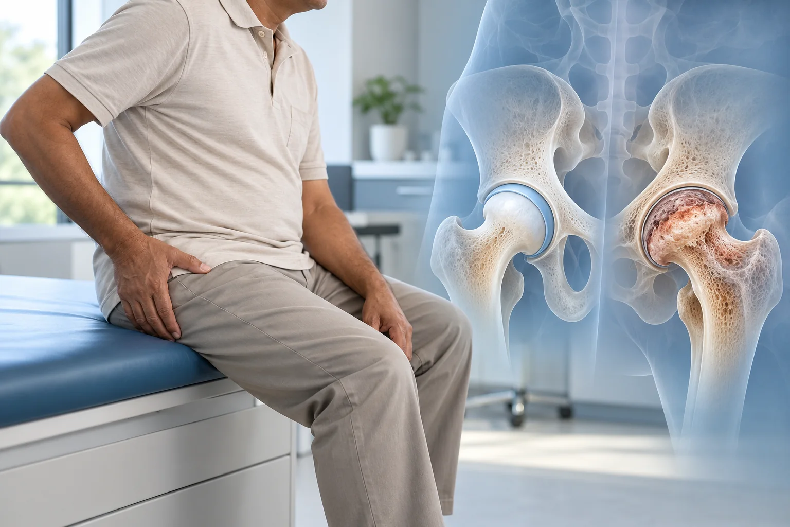
Your MRI report says avascular necrosis of the femoral head. You might have been told that part of your hip bone is damaged because it isn't receiving enough blood.
Then comes the difficult question: will you need a hip replacement?
You may also hear about a smaller operation called core decompression, which aims to preserve the natural hip. Why would one patient be offered that procedure while another is advised to consider replacing the joint?
With hip AVN, the answer often lies in something your scans reveal: whether the rounded upper end of the thighbone has started to lose its shape.
An early area of bone damage and a collapsed joint surface are not the same problem. Understanding the difference can help you make sense of your surgeon's recommendation.
Can Your Hip Be Preserved After an AVN Diagnosis?
Yes. Some patients with avascular necrosis can still be considered for joint-preserving treatment, particularly when the femoral head has not collapsed. Core decompression may be discussed at this stage, while hip replacement becomes more relevant when the bone has lost its shape and the joint is significantly damaged.
The decision is influenced by:
- Femoral-head shape: Is the rounded surface still intact?
- Size of the damaged area: How much bone is affected?
- Location: Is the damage beneath the part of the hip that carries most of your weight?
- Joint condition: Are the cartilage and hip socket still relatively preserved?
- Symptoms: How much pain, stiffness or difficulty walking do you experience?
These details matter more than choosing an operation based on age alone.
When significant collapse has occurred, patients may need to understand how hip replacement is assessed and planned before deciding whether replacing the damaged joint is appropriate.
The important point is that an AVN diagnosis does not automatically mean every patient needs the same operation.
What Is Actually Going Wrong Inside the Hip?
The hip works much like a ball sitting inside a socket.
The ball is the rounded top of your thighbone, known as the femoral head. The socket is part of your pelvis.
When both surfaces are healthy, their smooth cartilage coverings allow the joint to move with relatively little friction.
But the bone underneath that cartilage is living tissue. It needs blood to remain healthy.
Avascular necrosis, also called osteonecrosis, develops when the blood supply to a region of bone is disrupted or reduced enough for the affected bone tissue to die.
At first, the damage can remain hidden beneath an apparently normal joint surface.
You may still have a rounded femoral head on an X-ray, even though MRI has identified changes inside the bone.
Over time, the damaged region may weaken.
Walking and standing repeatedly place considerable pressure on the femoral head. If the weakened bone cannot withstand that load, tiny structural failures can develop beneath the joint surface.
Eventually, part of the rounded bone may flatten.
That is when the treatment discussion can change significantly.
AVN can occur after certain hip injuries and is associated with factors such as corticosteroid exposure, heavy alcohol use and some underlying medical conditions. Sometimes, no clear cause is found.
Why Can the X-Ray Look Normal When the MRI Shows AVN?
This is one of the more confusing parts of an early diagnosis.
You may have persistent groin pain, yet your X-ray report says there is no obvious joint damage.
That does not necessarily rule out AVN.
X-rays are useful for showing bone shape, fractures, joint-space changes and more advanced structural damage. They are less sensitive to early changes occurring inside the femoral head.
MRI can detect those changes before the bone's outer contour begins to flatten.
It also helps the surgeon see how far the affected area extends.
For example, an MRI showing a relatively small area of bone damage with an intact joint surface presents a different picture from one showing extensive disease beneath the weight-bearing region.
The US National Institute of Arthritis and Musculoskeletal and Skin Diseases (NIAMS) explains that MRI can detect osteonecrosis before changes become visible on conventional X-rays.
The MRI report is therefore useful, but the surgeon still needs to interpret the images alongside your symptoms and examination findings.
Three Scan Findings That Can Change the Treatment Discussion
Rather than focusing only on a stage number, it helps to understand what has physically happened to the femoral head.
1. Bone damage without collapse
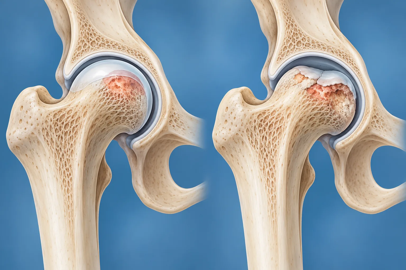
In the earliest stages, MRI may show an area of osteonecrosis while the rounded shape of the femoral head remains preserved.
The cartilage covering the joint may still be supported by an intact underlying surface.
This is the situation in which joint-preserving procedures are more likely to be considered.
However, not all early lesions behave the same way.
A large damaged region located directly beneath the main weight-bearing surface may have a greater risk of progression than a smaller lesion elsewhere.
2. A small fracture beneath the joint surface
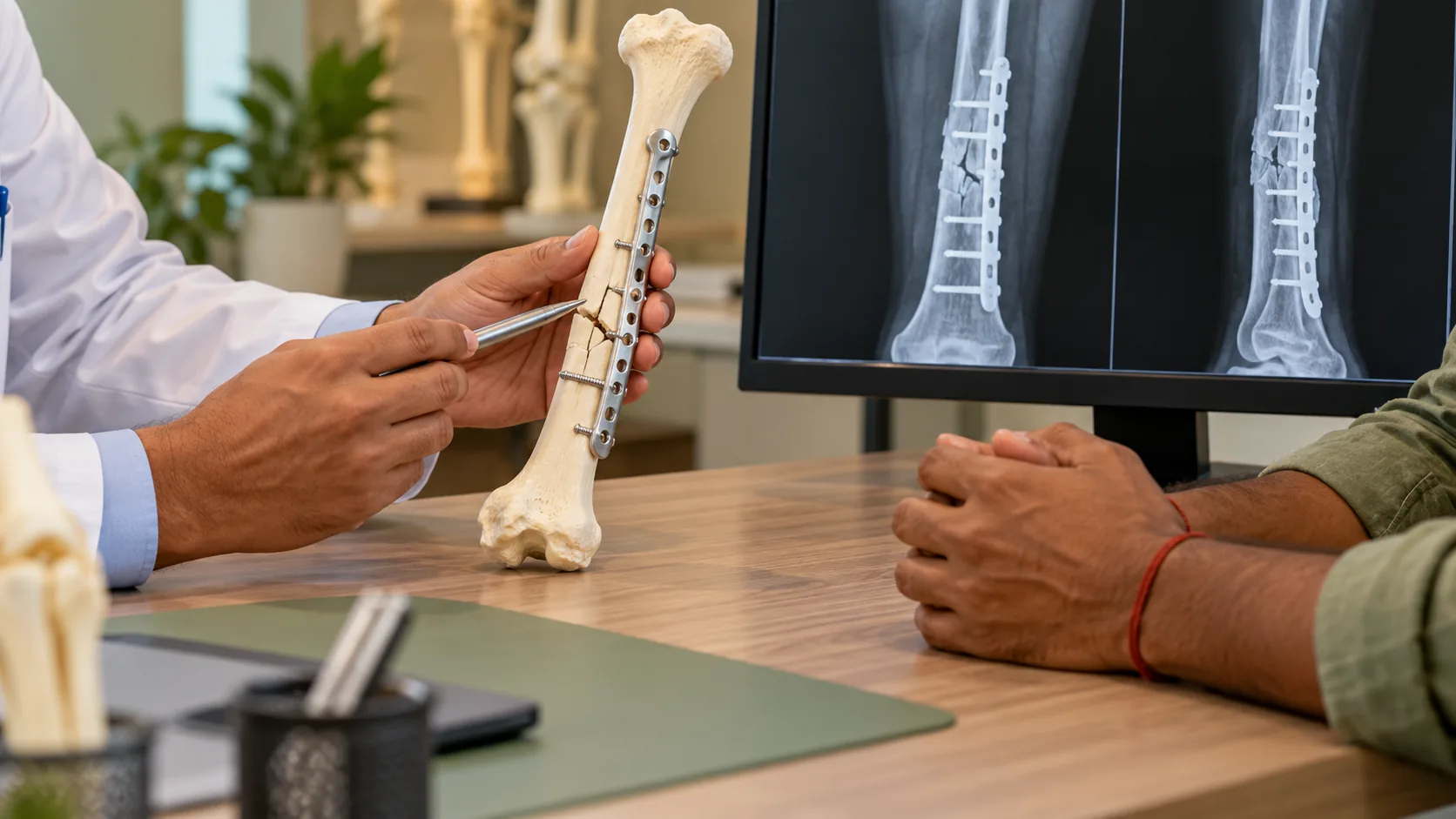
As the weakened bone begins to fail, a subchondral fracture may develop.
The term sounds complicated, but it simply means a fracture in the bone immediately underneath the joint cartilage.
Sometimes imaging shows a characteristic line associated with this change.
This finding suggests that the bone is losing some of its ability to support the surface above it.
It is an important distinction because the hip may still look reasonably rounded even as structural damage begins.
The surgeon needs to consider whether joint-preserving treatment remains appropriate and how likely further collapse may be.
3. Flattening or collapse of the femoral head
Once the weakened area gives way, part of the rounded femoral head can become flattened or irregular.
With more advanced disease, the joint surface may deteriorate further, and degenerative changes can develop in the socket.
At this point, the problem is no longer confined to damaged bone hidden beneath an intact surface.
The shape of the joint itself has changed.
That makes procedures intended to preserve the existing femoral head less likely to achieve their original goal.
These findings are related, but they are not interchangeable. An early MRI lesion, a fracture beneath the cartilage and established femoral-head collapse can lead to different recommendations.
When Might Core Decompression Help?
Core decompression is a surgical procedure designed to treat selected cases of early hip osteonecrosis.
During the operation, the surgeon creates one or more channels within the affected bone.
The procedure aims to reduce pressure inside the femoral head and support the biological processes involved in bone repair and blood-vessel formation.
In some cases, additional techniques, such as bone grafting, may be used.
The main advantage of considering core decompression early is that the natural joint surface may still be structurally preserved.
For example, a patient whose MRI identifies a limited area of AVN without collapse may be assessed for a joint-preserving approach.
But the operation isn't suitable for everyone with early disease.
The surgeon must consider how much bone is affected, where the damaged area sits and whether the femoral head is already beginning to fail structurally.
The American Academy of Orthopaedic Surgeons' guidance on osteonecrosis of the hip explains that core decompression generally has better results before the femoral head collapses.
It can help some patients avoid or delay replacement, but it cannot guarantee that AVN will stop progressing.
Some people eventually require hip replacement despite undergoing a joint-preserving procedure.
Why Might Hip Replacement Be More Suitable After Collapse?
Suppose the femoral head has become noticeably flattened.
The affected joint surface is no longer maintaining its normal rounded shape, and the hip may have developed associated cartilage damage or arthritis.
In this situation, creating channels inside the bone cannot reliably restore the original shape of the joint surface.
If pain and restricted movement are substantial, replacing the damaged joint may become a more appropriate surgical option.
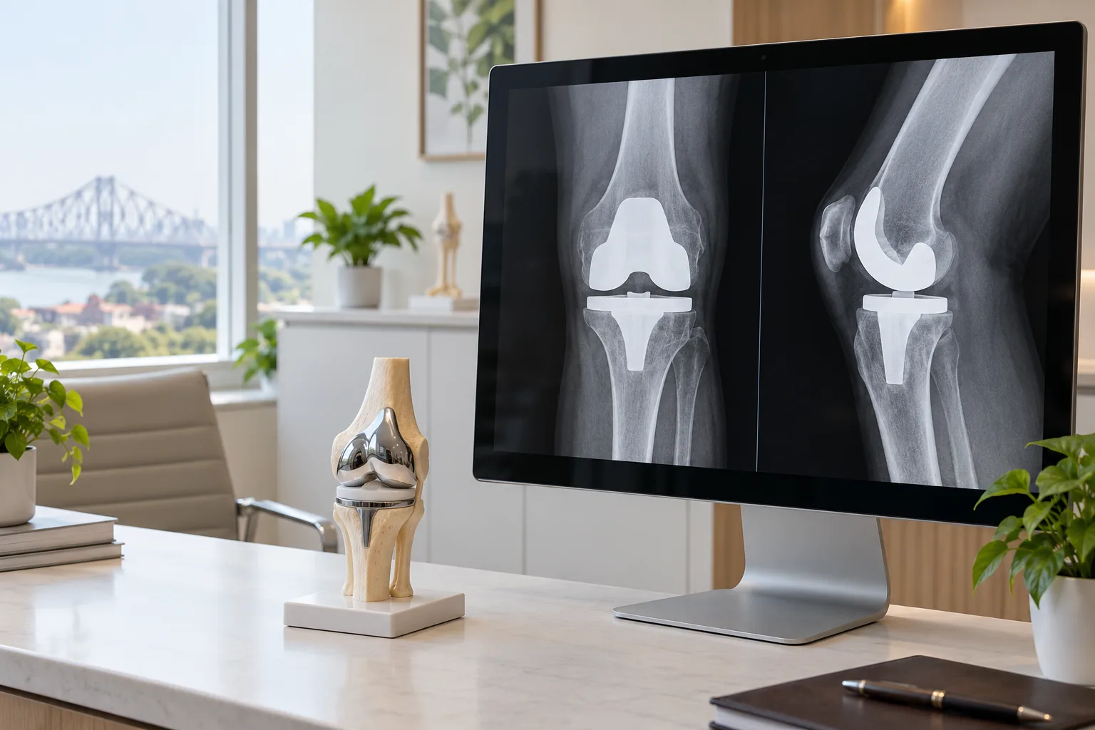
Total hip replacement involves removing the damaged femoral head and reconstructing the joint with artificial components.
A stem is placed into the thighbone and supports a new ball. The hip socket is prepared for an artificial cup and bearing surface.
The aim is to address the damaged articulation and improve pain and function.
This can make replacement a reasonable option for patients with advanced AVN, particularly when walking, climbing stairs, sitting comfortably or other routine activities have become difficult.
Still, collapse on an image does not automatically mean every patient must undergo immediate surgery.
The extent of joint damage, symptoms, overall health and personal treatment goals remain part of the decision.
Early AVN vs Advanced AVN: What Changes?
| What the surgeon assesses | Earlier AVN | More advanced AVN |
|---|---|---|
| Shape of femoral head | Usually remains rounded | May be flattened or collapsed |
| X-ray | May appear normal | Often shows structural changes |
| MRI | Detects early bone damage and its extent | Helps assess lesion extent and associated damage |
| Joint surface | Often structurally preserved | May be irregular or deteriorated |
| Core decompression | May be suitable for selected patients | Less likely to help after substantial collapse |
| Hip replacement | Usually not the first surgical option when preservation is feasible | More relevant when collapse and symptoms are significant |
| Main treatment objective | Preserve the existing joint where possible | Address the damaged joint surfaces |
This comparison explains the general direction of treatment, but it cannot determine the right operation for an individual patient.
A specialist may use formal staging systems, including Ficat or ARCO, to describe how far the condition has progressed.
The stage is useful, but the size and location of the lesion are also important.
Two people with the same reported stage may have very different areas of damage and levels of pain.
Does Being Young Mean You Should Avoid Hip Replacement?
A younger patient may have good reasons to prefer preserving the natural hip.
Hip replacement implants do not last indefinitely, and the possibility of future revision surgery matters when someone is expected to live with the implant for many decades.
However, being young does not make a badly collapsed femoral head suitable for core decompression.
Similarly, an older adult with limited early-stage disease should not be excluded from a joint-preserving discussion solely because of age.
What matters is the condition of the femoral head and whether the proposed operation is likely to provide a meaningful benefit.
The surgeon also considers general health, activity levels, bone condition and expectations.
Age influences planning. It should not replace an assessment of the actual hip damage.
Can Medicines, Rest or a Walking Aid Reverse AVN?
Non-surgical measures can help manage symptoms and reduce strain on the painful joint.
For example, a walking aid may make movement more comfortable by reducing the load passing through the affected hip.
Appropriate pain medication and adjustments to activity can also be useful.
But these measures do not reliably restore dead bone or reverse an established collapse.
They may form part of a treatment plan, particularly when surgery is not immediately indicated or while the diagnosis and stage are being assessed.
Addressing underlying risk factors also matters.
If you use corticosteroids for another medical condition, do not stop or change the medication without speaking to the doctor who prescribed it.
The cause of AVN and the treatment needed for any associated illness should be reviewed carefully.
A person with early AVN may require monitoring to detect progression even when symptoms are manageable.
How Does Recovery Differ Between Core Decompression and Hip Replacement?
The two procedures have different purposes, so their rehabilitation plans differ.
After core decompression
The natural femoral head remains in place.
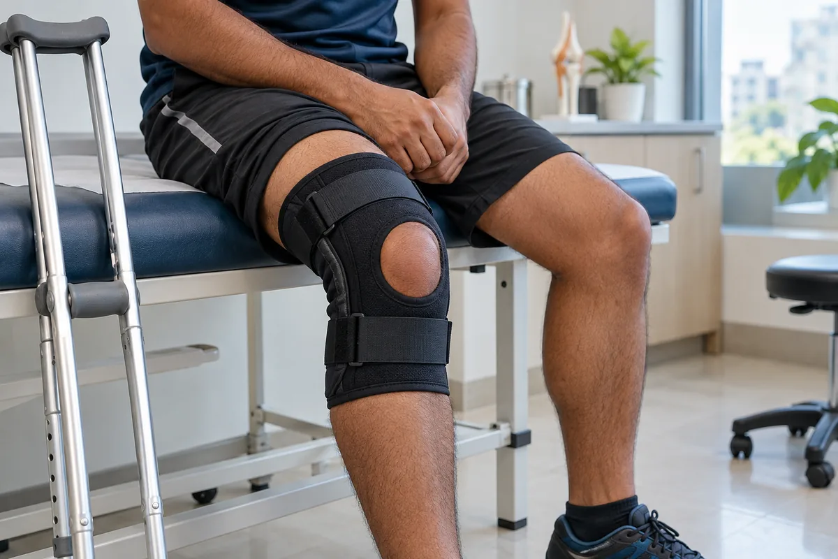
The treated bone needs time to recover, and the surgeon may recommend crutches or another walking aid with restrictions on how much weight can be placed through the hip.
Follow-up assessments help determine whether the affected area is healing and whether the joint remains structurally stable.
The main uncertainty is whether the procedure will provide enough benefit to prevent further deterioration.
Recovery can take several months, and progression may still occur.
After hip replacement
Rehabilitation focuses on recovering from the operation and gradually improving movement, balance and strength with the artificial joint.
Depending on the patient's condition and surgical plan, walking with assistance may begin relatively early.
Progress continues over the following weeks and months.
Possible complications include infection, blood clots, dislocation, leg-length differences, nerve injury and implant-related problems.
Although hip replacement can substantially improve symptoms in appropriately selected patients, the degree and speed of improvement vary.
Neither procedure should be described as a guaranteed cure.
What Should You Understand Before Making a Decision?
If your MRI report says AVN, try to understand more than the diagnosis itself.
The most useful details concern the condition of the femoral head.
Has the rounded surface remained intact? Is there a fracture just beneath the cartilage? Has the bone started to flatten? How much of the weight-bearing area is affected?
These findings help explain why core decompression may be discussed in one patient while replacement is recommended for another.
A surgeon should also explain the expected benefits, limitations, risks and rehabilitation requirements of the proposed operation.
If a joint-preserving procedure is being considered, understand the possibility that AVN may still progress.
If replacement is recommended, understand what damage makes preservation less suitable.
You should have enough information to make a decision without feeling that a particular operation is inevitable simply because AVN has been diagnosed.
When Should You Consult a Hip Specialist in Kolkata?
Persistent groin pain, limping, stiffness or difficulty bearing weight deserves assessment, particularly when symptoms are getting worse.
People already diagnosed with AVN should also attend recommended follow-up appointments, because disease progression may affect treatment options.
At HealMyBones, Dr. Manoj Kumar Khemani provides orthopaedic and hip replacement consultations in Kolkata.
HealMyBones encourages patients to understand their diagnosis, available treatments and individual medical findings before making decisions about surgery.
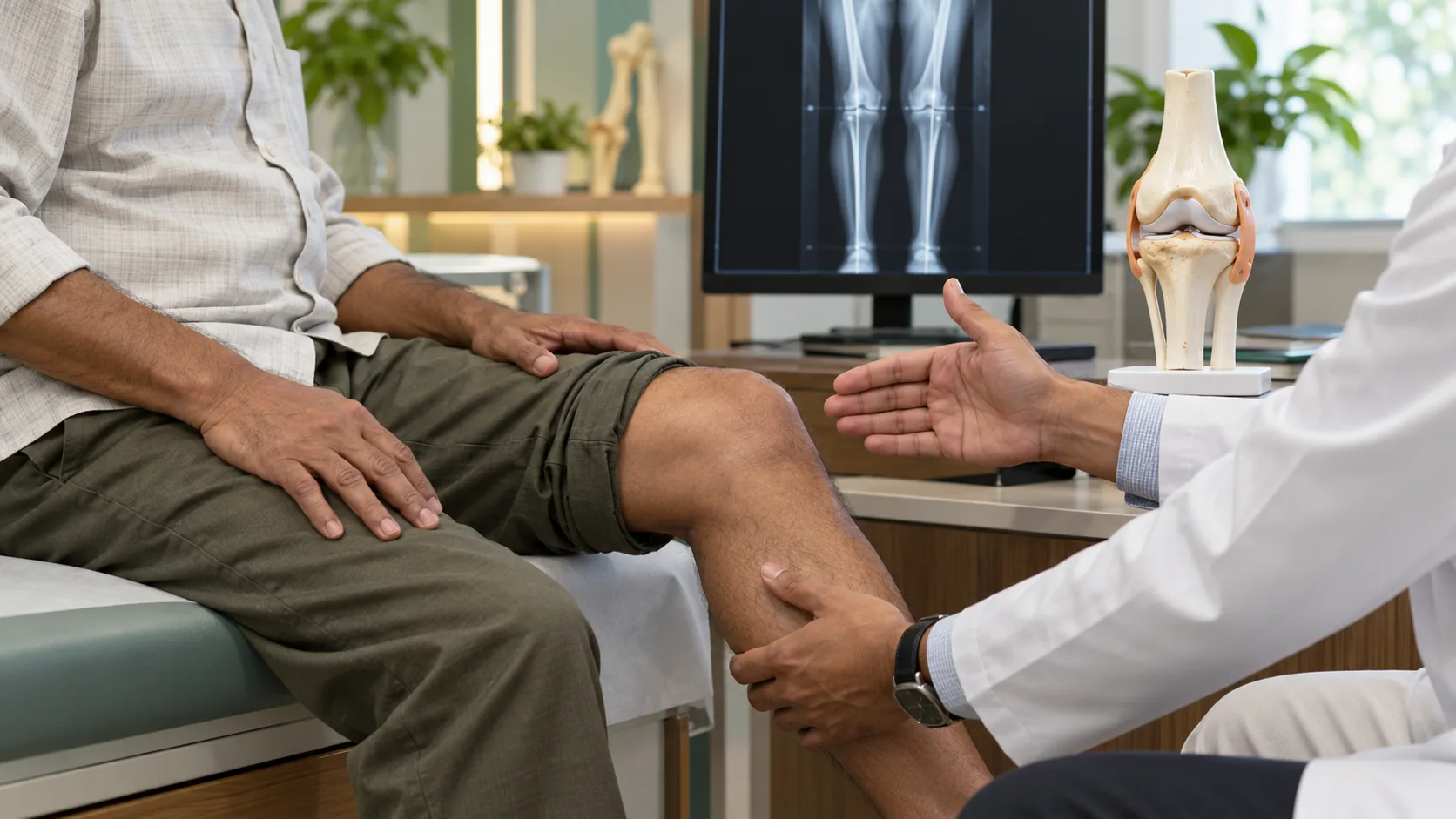
For someone researching avascular necrosis hip treatment in Kolkata, the most useful consultation is one that reviews the imaging and explains whether the joint surface is still suitable for preservation.
Sudden severe pain or a new inability to bear weight, especially after a fall, requires urgent medical assessment.
FAQ
Can hip AVN affect both hips at the same time?
Yes, avascular necrosis can affect both hips, even when pain is initially felt on only one side. Bilateral involvement is more common with certain non-traumatic causes, although severity may differ between hips. An orthopaedic specialist can determine whether assessment or imaging of the other hip is appropriate.
Does every person with Stage 2 hip AVN need core decompression?
No, Stage 2 AVN does not automatically mean core decompression is necessary. Lesion size, location, symptoms and the absence of collapse influence whether the procedure is suitable. A surgeon should review the actual MRI images rather than recommend an operation from the stage number alone.
Can hip AVN get worse even when the pain has reduced?
Yes, symptoms can fluctuate, and less pain does not reliably prove that the affected bone has healed. Some lesions may continue to progress structurally despite periods of symptom improvement. Follow-up imaging and examination should be guided by the treating specialist.
If core decompression fails, can hip replacement still be performed?
Yes, hip replacement may remain an option if AVN progresses after core decompression. The surgeon must consider the remaining bone condition, prior procedures, symptoms and any associated joint damage. Patients should discuss the possibility of future surgery when considering a joint-preserving procedure.
Your Scan Matters More Than the Name of the Operation
Core decompression and hip replacement are not interchangeable treatments.
One aims to preserve the natural femoral head while it still has enough structural integrity. The other replaces damaged joint surfaces when keeping the original joint may no longer provide adequate function.
The decision begins with understanding what AVN has done to the bone.
If your MRI or X-ray shows avascular necrosis, ask for a clear explanation of whether the femoral head is intact, beginning to fracture beneath the surface or already collapsed.
That discussion can help you understand why a particular treatment is being recommended—and what the realistic alternatives may be.
Medical Disclaimer
This article is intended for general education only and does not constitute individual medical advice, diagnosis or a recommendation for surgery.
Avascular necrosis varies in severity, and treatment decisions depend on medical history, symptoms, physical examination and appropriate imaging. Core decompression and hip replacement carry risks, and neither procedure guarantees pain relief, prevention of further bone damage or restoration of normal hip function.
Consult a qualified orthopaedic professional for personalised advice. Seek urgent medical care for sudden severe hip pain or an inability to bear weight, particularly after an injury.