Dr. MANOJ KUMAR KHEMANI
(Specialist Orthopaedic Surgeon)
Categories
Treatments

Shoulder Replacement

What is shoulder replacement?

Shoulder joint is a complex joint designed to provide extreme flexibility. However due to this flexibility there is some compromise in the stability of the joint.
Just like hip and knee, the shoulder joint is also prone to arthritis, although in much less number of people.

Since it’s not a weight bearing joint, a little amount of arthritis in the shoulder joint is usually not very disabling. Some patients do have arthritis of the shoulder to such an extent that it causes a lot of trouble in their day to day activities.

When is shoulder replacement done?

Shoulder replacement is an excellent choice of treatment in severely damaged shoulder joint where nonsurgical treatments like medications and activity changes are no longer helpful for relieving pain.

shoulder replacement

Conditions where this operation is done:

  • Osteoarthritis
  • Rheumatoid arthritis
  • Post-Traumatic arthritis
  • Rotator Cuff Tear Arthropathy
  • Avascular Necrosis (Osteonecrosis)
  • Irreparable fractures

Types of shoulder replacement:

Shoulder replacement are of these types:

  • Total shoulder replacement
  • Reverse shoulder replacement
  • Stemmed Hemiarthroplasty
  • Resurfacing Hemiarthroplasty

Categories
Treatments

Types of Arthritis

What are the various types of arthritis?

Arthritis encompasses several conditions characterized by joint pain as a primary symptom. Arthritis can be of many different types. When an individual is diagnosed with “arthritis,” it typically indicates they have one of the following diseases:

1. Osteoarthritis
2. Rheumatoid arthritis
3. Gout and pseudo-gout
4. Ankylosing spondylitis
5. Septic arthritis
6. Juvenile idiopathic arthritis
7. Still’s disease

Types of arthritis

Additionally, joint pain can serve as a symptom of other diseases, where arthritis is considered secondary. These include conditions such as:

– Psoriasis (Psoriatic arthritis)
– Reactive arthritis
– Ehlers-Danlos Syndrome
– Haemochromatosis
– Hepatitis
– Lyme disease
– Sjogren’s disease
– Hashimoto’s Thyroiditis
– Inflammatory bowel disease (including Crohn’s disease and ulcerative colitis)
– Henoch-Schönlein purpura
– Hyperimmunoglobulinemia D with recurrent fever
– Sarcoidosis
– Whipple’s disease
– TNF receptor-associated periodic syndrome
– Wegener’s granulomatosis (and various other vasculitis syndromes)
– Familial Mediterranean fever
– Systemic lupus erythematosus
– Post Traumatic Arthritis

Undifferentiated arthritis refers to a type that does not fit into well-known clinical disease categories, possibly representing an early stage of a definite rheumatic disease.

Can arthritis result from injury?

Permanent damage to joint cartilage and ligaments due to injury can lead to post-traumatic osteoarthritis, causing joint failure over time. Early treatment if joint injuries aim to minimize this damage.

Can an individual have more than one type of arthritis?

Generally, individuals tend to experience only one type of arthritis. Nevertheless, in prolonged cases, there is a potential progression towards secondary osteoarthritis. On occasion, a unique scenario arises where the simultaneous presence of two types of arthritis, such as rheumatoid arthritis and gout, may manifest.

How is arthritis diagnosed, and what are the treatment options?

Despite thorough investigations, identifying the precise type of arthritis can be challenging. Subsequently, treatment initiates based on the doctor’s clinical judgment and supportive investigations, with modifications made according to the patient’s response. Although arthritis lacks a permanent cure, a diverse array of treatments exists, encompassing anti-inflammatory medications, physiotherapy, exercises, and the application of hot or cold compresses for joint relief.

In the event of a diagnosis of secondary arthritis, treatment aims to address the underlying condition. Notably, in certain cases, healthcare professionals may recommend injections directly into the affected joint as part of the therapeutic approach.

Some major joints like hip, knee, shoulder and elbow, when severely damaged, can be replaced with artificial components with help of replacement surgeries (also called arthroplasty).

What are Knee and Hip Replacements?

Knee Replacement is a surgical procedure performed in severe osteoarthritic knees to alleviate pain and restore function. Similarly, Hip Replacement is undertaken in cases of hip fractures or damage to restore hip function and manage pain.

Dr. Manoj Kumar Khemani has more than 15 years experience in diagnosing and treating various forms of arthritis. You can contact him using this contact form.


Categories
Treatments

What is Arthritis?


Arthritis is a disease of joints in our body. It means “inflammation of the joint” . Maybe one or more. The commonest complain in a patient of arthritis is joint pain.

Arthritis is not a single disease – it is a term that covers over 100 medical conditions.

Osteoarthritis (OA) is the most common form of arthritis and generally affects elderly patients. This disease affects cartilage, the tissue that cushions and protects the ends of bones in a joint.

Another common form of arthritis is Rheumatoid Arthritis. This is an auto-immune disease in which the body’s immune system (the body’s way of fighting infection) attacks healthy joints, tissues, and organs. Occurring most often in women of childbearing age (18-44), this disease inflames the lining (or synovium) of joints.

Gout or Uric acid arthritis is also common in Indian people. It occurs due to deposition of uric acid crystals in the joint, these crystals irritate and scratch the tender joints to cause pain, inflammation and finally erosion.

Some forms of arthritis can affect people at a very early age e.g. Juvenile Rheumatoid Arthritis.

No matter what the type of arthritis it is, it always causes discomfort.

Some of the common types of arthritis are listed below:

  • Osteoarthritis
  • Rheumatoid Arthritis
  • Gout and Pseudo-gout
  • Psoriatic Arthritis
  • Juvenile Arthritis
  • Ankylosing Spondylitis
  • Polymyalgia Rheumatica
  • Septic arthritis
  • Systemic Lupus Erythematous
  • Post Traumatic Arthritis

Dr Manoj Kumar Khemani best ortho Knee replacmentDr. Manoj Kumar Khemani has over 15 years experience in treating advanced arthritis. He can monitor your condition, adjust your treatment plan as needed, and provide guidance on the best arthritis treatment in Kolkata.

Categories
Treatments

Vitamin D

Vitamin D: A gift from the sun.

Vitamin D is one vitamin which is produced in our body by direct exposure to the sun. The vitamin D formed in the skin is processed in our liver and then kidneys to finally form produce the active form of Vitamin D [1,25(OH)2D].

What is the function vitamin D?

Vitamin D plays an important role in absorption of calcium from our food which we eat. It is also needed for bone growth and bone remodeling. Recent studies have shown that it also has role in our neuromuscular and immune function, reduction of inflammation and cell growth.

Research suggests that vitamin D could play a role in the prevention and treatment of a number of different conditions, including type1 and type 2 diabetes, hypertension, glucose intolerance, and multiple sclerosis.

What causes it’s deficiency?

No food items contain adequate vitamin D. Therefore to get sufficient D you need good amount of strong sunlight. Lack of sun exposure is the primary cause of it’s deficiency. Contrary to popular belief early morning sunlight is not sufficient. You need exposure to sun at noon. That’s when the UV rays are strongest.

Other reasons are, insufficient absorption of dietary supplements of vitamin D due to intestinal diseases and failure of kidneys to convert it into active form.However prolonged UV ray exposure has it’s own hazards (including skin cancer). That’s why most doctors rely on supplements rather than prescribing sun exposure.

Effects of vitamin D deficiency:

Vitamin D deficiency in children can cause severe deformities in bone. This disease is known as Rickets. Common presentation of rickets is bow legs. If diagnosed early this condition is reversible by giving adequate vitamin D in diet. As the bone matures the effects become permanent.

In adults the deficiency is known as Osteomalacia. It is characterized by weak bones. In prolonged cases there can be bone pain and muscle fatigue.

Vitamin D deficiency has also been linked with cardiovascular diseases, cancer, autoimmune diseases, infections and some metabolic disorders.

Diagnosis:

It can be easily diagnosed by blood test estimating 25(OH)D3.

Treatment:

Treatment is aimed at correcting the vitamin D levels in the body. Vitamin D supplements are now available in many forms including tablets, capsules, powder, liquid and injection.

In most patients weekly high dose is given for a few weeks to rebuild vitamin D store quickly. Following which a daily or monthly maintenance dose is recommended.

What will happen if too much vitamin D is taken?

Vitamin D is a fat soluble vitamin and is stored in our body. Unlike some other vitamins very high dose of vitamin D for long period has been associated with significant risk factors. Hypercalcemia and deposition of calcium in kidneys and other tissues can occur.

High dose of vitamin D must always be taken under guidance of a doctor.


Categories
Treatments

11 Myths about Osteoporosis

Do you know that much of what we’re told about bone health is actually a myth? In reality, there’s a lot you can do to build bone strength, prevent osteoporosis and reduce fracture risk. Let’s brush our knowledge…

Myth 1: You cannot have osteoporosis if you take regular calcium.

Yes, calcium is important, but it’s a myth that simply taking a high amount of calcium will guarantee bone health. To protect your bones, you need many additional essential nutrients, not just calcium. In fact, you need some of those nutrients just to get any benefits of calcium. For example, without enough vitamin D, your body only absorbs about 10-15% of the calcium from your diet, but when you take enough, the absorption rate jumps to 30-40%. Other critical nutrients for bone health are magnesium, vitamin K, vitamin C, boron, strontium etc.

Modern lifestyle prevents us from getting adequate sunlight, which is important for synthesis of vit. D in our body.

Myth 2: Osteoporosis is normal… as your bones age they should get weak.

Bone loss, even osteoporosis can affect you in your 30’s and 40’s. One of the most dangerous bone health myths is that osteoporosis is inevitable as we age. Remember, osteoporosis is not normal. It’s a disease. While there are some fixed risk factors — such as our age and gender — you can control many of the risk factors that lead to excessive bone loss, osteoporosis and fracture. The truth is, you can have strong bones at any age.

Myth 3: A diagnosis of osteoporosis means you’ll suffer a fracture.

Many people with osteoporosis may not have fracture. What’s more, many people who have normal bone density do experience fractures. Osteoporosis does increase your fracture risk.To identify your personal fracture risk, you can use this WHO fracture risk assessment tool.

Myth 4: Lack of estrogen causes osteoporosis – it’s a woman’s problem.

We hear all the time that osteoporosis is a “women’s disease” and men don’t really need to worry about it. This may stem from the long-standing belief that low estrogen levels cause bone loss. But in many countries, women maintain healthy bones for life — even though they experience the same lower estrogen levels with menopause the rest of us do. So while estrogen may play a role in osteoporosis, it’s certainly not the major cause. And unfortunately, men get osteoporosis too!

Myth 5: Osteopenia leads to osteoporosis.

A diagnosis of osteopenia means you have a state of relatively low bone mass, compared to the standard. For many women this may be only in one area, not necessarily throughout your body. And it doesn’t always mean that you’re currently losing bone. Your bones are alive. It’s never too late to build bone because it’s living tissue that constantly repairs itself.

Myth 6: You don’t need to worry about osteoporosis until menopause.

Bone loss — even osteoporosis — can be secretly affecting you in your 20s, 30s and 40s. We normally achieve peak bone mass in our 20s and then begin to lose it, some of us more quickly than others. The earliest type of bone loss takes place for women who are thin, have celiac disease, suffer from irregular menstrual cycles or poor nutrition, or use steroid drugs.

Myth 7: There’s nothing you can do once you have osteoporosis other than take a drug.

You can’t be more wrong. The natural way to bone health combines nutrition, physical activity and fall prevention. Next comes assessing and treating the underlying causes of compromised bone health. Finally, medicines help you regain your bone mass quickly. In a nutshell it’s lifestyle modification.

Myth 8: Osteoporosis is common all over the world.

Osteoporotic fracture rates vary greatly around the world. Unfortunately in India we don’t have sufficient data on osteoporosis due to the lack of awareness. It’s clear that certain lifestyle factors play major roles in bone loss, including:

  • Stress and anxiety
  • Lack of exercise
  • High caffeine intake
  • Poor nutrition
  • Use of certain medicines (e.g. steroids, anti-epliptics)
  • Cigarette smoking
  • Alcohol

Myth 9: Osteoporosis isn’t linked to other health issues.

More and more, research appears to indicate that there’s a link between the existence of osteoporosis and other diseases. When you build your bones, you’re likely building a healthier, stronger body and improving metabolic fitness, muscle strength, blood pressure regulation and cardiovascular health — all at the same time. Conversely people with known medical conditions are at a higher risk of osteoporosis.

Myth 10: There aren’t any signs or symptoms of bone loss.

While many people don’t realize they have a bone issue until they fracture, there are sometimes signs and symptoms of bone loss. These include receding gums; decreased grip strength; weak and brittle fingernails; cramps, muscle aches and bone pain; height loss and low overall fitness. These are often late and variable and you should not solely rely on them.

By knowing these facts about bone loss, osteoporosis and fracture, you can take the steps to have better bones now and for life.

Myth 11: Osteoporosis and Osteoarthritis are same things.

While Osteoporosis is loss in bone density, osteoarthritis is degeneration of joints. They are completely different diseases and should not be confused together.

Who Should Get Screened for Bone Loss

In general, people who should be screened for osteoporosis include:

  • Women over age 50
  • Women who have more than one risk factor (for example: a family history of osteoporosis, post menopause, low body weight etc.)
  • Post-menopausal women who have had a fracture

Since bone loss begins without any noticeable symptoms, screening should ideally occur before you have reason to worry.

You can use this WHO international Fracture Risk Assessment tool to find out your true risk.

You can also visit me in my clinic. See my clinic schedule.

Categories
Treatments

Osteoarthritis

A Normal Joint

A joint is where two or more bones meet. A joint allows your bones to move freely but within controlled limits.

To prevent friction and erosion of bone the ends of your bones are coated with a layer of tough, slippery tissue called cartilage. Cartilage acts a bit like a shock absorber to spread the load more evenly across your joint. Also most joints have a small amount of synovial fluid in them which acts like a lubricant. Your knees have extra rings of cartilage between the bones. These are called menisci.

Although fractures might not be entirely preventable there are ways to minimize your fracture risk. One way is by building stronger bones and keeping them healthy.

What is Osteoarthritis?

It is a medical condition that affects your joints. In osteoarthritis (Sometimes called OA), there is gradual degradation of the joint cartilage due to wear and tear. A variety of causes like hereditary, developmental, mechanical and metabolic – initiate a process of cartilage degradation. As this process continues, the cartilage loses its ability to act as shock absorber. At some places there may be so much wear that the underlying bone becomes exposed and starts rubbing against each other. This causes pain and stiffness and its difficult to use the joint. There is also loss of structure and function of the supporting ligaments and muscles , causing instability.

It’s the most common type of arthritis commonly affecting the hip, knee and spine. However any joint can be affected. The chance of osteoarthritis in a joint increases with age. Most people above the age of 60 get osteoarthritis to some degree.

What are the signs and symptoms ?

Pain in the affected joint is the commonest symptom. Joint pain of osteoarthritis is usually worse later in the day. There can be swelling, warmth, and creaking noise in the affected joints. Pain and stiffness of the joints can also occur after long periods of inactivity (for example, sitting in a theater, getting up from bed in the morning etc.). In severe osteoarthritis, there can be pain even at rest or pain with limited motion and walking can become extremely difficult.

OA in the fingers and hands can limit fine movements. In the spine it can cause persistent neck or back pain.

Diagnosis of osteoarthritis

Diagnosis is straightforward based on :

  • Your description of symptoms
  • Physical examination by your physician
  • X-Rays
  • MRI

Sometimes your doctor may prescribe blood tests to make sure you don’t have other type of arthritis.

Types of Osteoarthritis

1. Primary : Occurring due to normal wear and tear of a joint.

2. Secondary : Due to an underlying cause like —

  • Inflammatory diseases / arthritis, such as rheumatoid arthritis
  • Previous Injury
  • Obesity
  • Inactivity or sedentary lifestyle
  • Genetic

Treatment of osteoarthritis

Early Stages – (Grade 1)

Osteoarthritis treatment depends on the grade of disease progression. In early stages the treatment comprises of a combination of-

  • Exercises
  • Physical therapy
  • Nutritional supplements
  • Weight loss
  • Medicines
  • Hot and cold compress
  • Application of pain reliving ointments / gels / oils

Intermediate Stages – (Grade 2, Grade 3)

With the progress of the disease your doctor may prescribe injection of medications into the joint. These can be either steroid injections or specialized gel formulations.

Late Stages – (Grade 4)

Surgery may be helpful to relieve pain when other treatment options have not been effective.

Both Hip replacement surgery and Knee replacement surgery have shown excellent long term results in treatment of severe arthritis.

The type of treatment will depend on several factors, including your age, occupation, activities , overall health, medical history, location of your osteoarthritis, and severity of the condition.

Categories
Treatments

Uric Acid Arthritis

What is Gout?

In normal people some amount of uric acid is always found in the blood. This remains in dissolved form. In people suffering from gout there is excess uric acid in the blood which crystallizes in one or more joints in the body. These excess needle shaped crystals of uric acid cause immense irritation in the joint. The result is pain and swelling of the joint.

How is uric acid formed?

Uric acid is formed from a class of protein called purines. It is a natural breakdown product of this protein. This conversion is done in our liver and small intestine. This uric acid is excreted out of our body through the kidneys. In normal people there is good balance between formation and excretion of uric acid.

What are the signs and symptoms ?

Sudden onset of severe pain and swelling in one or more joint is hallmark of gout. Usually this occurs overnight. Common joints involved in gout are first meta-carpo phalangeal joint (base of great toe), knee joint and ankle joint.

This pain usually subsides in 7-10 days. This pain might stay longer in repeated attacks of gout (chronic gout). Pain gets dramatically relieved with pain-killers.

It is more frequently seen in middle aged men and in some women after menopause.

Diagnosis of gout

Diagnosis can be made by the typical clinical presentation.

Confirmation of diagnosis is done by one or more investigations:

  • Increased uric acid in the blood
  • Polarized microscopy
  • X-Rays

Sometimes more blood tests are done to check if you have other type of arthritis.

Do I have gout?

You might be suffering from gout if you have the above mentioned symptoms. It’s very important to consult an orthopedic surgeon if you are having these symptoms as delay in treatment can cause permanent damage to your joints.

Some people have high levels of uric acid but don’t have any symptoms. These people are said to have hyperuricemia. Although it’s a risk factor, it’s not always necessary that the person will have an attack of gout.

What should I do if I have an attack of gout?

  • Rest.
  • Drink plenty of water.
  • Apply warm water over the affected area.
  • Visit an orthopaedic surgeon nearby.
  • Avoid high protein diet and alcohol.

What are the risk factors?

Risk factors for an attack of gout are:

  • High protein diet containing purines.
  • Alcohol intake
  • Stress
  • Dehydration
  • Family history of gout
  • High levels of uric acid in blood
  • Beverages sweetened with fruit sugar (fructose)

My uric acid is normal, still I have gout like pain

In some people the serum uric acid levels might be normal during an attack of gout. This is due to the fact that most of the uric acid has already crystallized into the joint. It may also be due to some other type of arthritis. Your doctor can usually differentiate between the different arthritis but sometimes it’s difficult and requires additional tests.

Deposition of calcium pyrophosphate can have similar symptoms as gout. This is condition called pseudo gout. Serum uric acid is normal in this case.

Is there a permanent cure for gout?

There is no permanent cure for gout. There are medicines which help to lower the uric acid levels and help to prevent gout attacks.

Treatment of Gout

Gout is treated with :

  • Diet control
  • Uric acid lowering medicines
  • NSAIDs

Food items to avoid in Gout

Gout is treated with :

  • Alcohol
  • Sea food
  • Red meat,
  • Organ meat (liver, kidney)
  • fish, poultry
  • Spinach
  • High-fructose corn syrup (HCFS)
  • soya bean
Categories
Treatments

Rheumatoid Arthritis

What is Rheumatoid Arthritis (RA)?

Rheumatoid arthritis one of the commonest type of inflammatory arthritis. In this disease our immune system behaves strangely and starts attacking the tissues of the joints (autoimmune disease). The result is that there is marked swelling and pain in the joints followed by irreversible destruction of the joint.

Women are more affected than men. The disease most often begins between the ages of 30 and 50. However, RA can start at any age.

Clinical presentation of Rheumatoid Arthritis.

Patients of RA have joint pain, stiffness, swelling and decreased movement of the joints. This pain is chronic in nature with episodes of aggravation and remissions. Small joints in the hands and feet are most commonly affected. Rarely RA can affect organs, such as eyes, skin or lungs.

Over a period of the time the pain gets worse and characteristic deformities start appearing in the joints. There is marked stiffness when getting up from bed in the morning.

Other symptoms seen are:

  • Marked weakness
  • Low grade fever
  • Loss of appetite
  • Polyarthralgia
  • Firm lumps, called rheumatoid nodules, which grow beneath the skin in places such as the elbow and hands
  • Anemia

It is commonly seen in middle aged females but can affect people of all ages.

The cause of RA is not known. There is evidence that autoimmune conditions run in families. For instance, certain genes that you are born with may make you more likely to get RA.

Diagnosis of RA.

Diagnosis of RA.

Confirmation of diagnosis is done by one or more investigations:

  • Increased RA factor in the blood
  • Increased ESR and CRP
  • Presence of anti-CCP in blood
  • X-Rays

Sometimes more blood tests are done to check if you have other type of arthritis.

Treatment of RA

In the last 30 years there has been significant improvement in the treatment of RA. Current treatments give most patients good or excellent relief of symptoms and let them keep functioning at, or near, normal levels. Treatment is started with a class of medicines called DMARD’s. These medicines help modulate to immune system to minimize inflammatory response. These may be combined with low dose corticosteroids.

Historically gold containing medicines have been used to control RA. However these have been largely abandoned due to potential side effects.

Newer medicines called “Biologic agents” have shown promise in cases where traditional medicines have failed.

In some patients injection of corticosteroids in the joint helps to reduce the pain.

Patients with advanced arthritis of hip, knee, shoulder and elbow are treated with joint replacement surgery.

Is there a permanent cure for RA?

There is no permanent cure for RA. Treatment is aimed at controlling the pain and inflammation, and minimizing joint destruction. Early diagnosis and treatment is key to successful outcome. Once there is permanent damage to the joint, complete pain relief is difficult.

What should I do if I have RA?

  • Take medicines regularly
  • Do regular exercise
  • Drink plenty of water.
  • Apply ice packs over the affected area.
  • Regular checkup with an orthopaedic surgeon or rheumatologist.
  • Swimming helps to keep the joints mobile.

Related Article:

Winter and Arthritis: 7 Tips for Pain Relief

Categories
Treatments

Frozen Shoulder (Adhesive Capsulitis)

What is Frozen shoulder?

It’s a medical condition characterized by severely reduced range of motion of the shoulder.

Also known as adhesive capsulitis.

Usually occurs in people over 40 years of age.

If you suffer from diabetes, heart disease, connective tissue disorders or have had a stroke or a severe injury, then you are more at risk of getting frozen shoulder.

What causes Frozen Shoulder Syndrome?

The exact cause is unknown and there are several theories about its cause. Many physicians believe the causes are linked to myo-fascial trigger points associated with the muscles of the shoulder. Slowly the muscles become less flexible and the corresponding shoulder joint gradually becomes locked into place over a period of time.

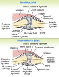
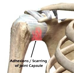
Frozen shoulder may also occur due to inflammation of the joint lining which causes scar tissue to form around the joint. This scar tissue restricts movement in the shoulder causing it to “freeze” and can be very painful.

It’s also believed incomplete healing of a previous shoulder injury can lead to frozen shoulder as the initial injury creates a buildup of scar tissue and inflammation in the shoulder joint.

The main symptoms are:

  • Decreased motion of the shoulder
  • Stiffness
  • Pain
  • Night cramps

Frozen Shoulder usually starts with pain. This pain prevents the patient from moving their arm. Lack of movement leads to stiffness and eventually less motion. Over time, you become unable to do movements such as reaching over your head or behind you.

If you have any of the above symptoms you should immediately see an Orthopedic surgeon.

Depending on your condition your doctor can prescribe you the following treatment options:

Conservative treatment options:

  • Anti-inflammatory medication
  • Physical therapy (physiotherapy)
  • Steroid injections

In some cases, it may only take a few weeks to see progress, but in others, it may take as long as 6 – 9 months for complete recovery. Physical therapy is required in some cases and it can be intense, needing to be done every day.

Surgical treatment options:

Surgery is recommended if nonsurgical treatment is not effective.

In cases of moderate to severe frozen shoulder only manipulation under anesthesia may be attempted. The patient is put to sleep using general anaesthesia and the shoulder is manipulated to its full range using controlled force. This procedure takes only 10-15 minutes.

In some severe cases shoulder arthroscopy is done under anesthesia. During this surgery, the scar tissue is released by bringing the shoulder through a full range of motion. Arthroscopic surgery can also be used to cut the tight ligaments and remove the scar tissue from the shoulder. After surgery, the patient may receive pain blocks so they can participate in physical therapy.

Whatever the choice of treatment, physical therapy is very important for full recovery.

Prognosis

Treatment with only physical therapy and non steroidal anti-inflammatory drugs will usually restore motion and function of the shoulder within 6 months. Recovery is faster if treatment is started early.

After surgery restores motion, patients must continue physical therapy for several weeks or months to prevent the frozen shoulder from returning. Treatment may fail if you cannot keep up with physical therapy. Surgery has the potential to speed recovery and improvement in range of motion.

Prevention of frozen shoulder

One of the most common causes of frozen shoulder is the immobility that may result during recovery from a shoulder injury, broken arm or a stroke.

If you have had an injury that makes it difficult to move your shoulder, talk to your doctor about what exercises would be best to maintain the range of motion in your shoulder joint before it reaches the frozen shoulder stage. Start a rehab protocol as early as possible.

If you haven’t had a recent shoulder injury and are still experiencing shoulder pain and stiffness that limits your range of motion for an extended period of time, once again contact your doctor for an evaluation. Early treatment and physical therapy can lead to fewer complications and quicker recovery.